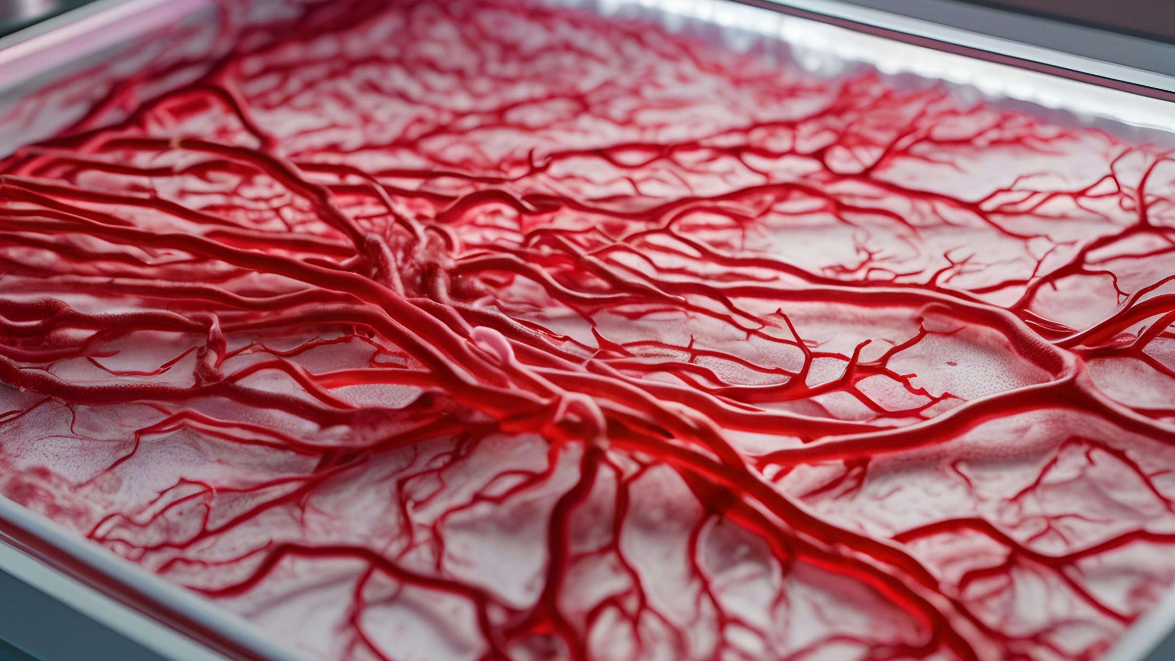
We can 3D print blood vessels now
3D printing isn’t new in the organ transplant and manufacturing space. But creating true-to-life blood vessel networks has long eluded researchers. Until now.

The story: A Harvard research team has 3D printed blood vessels that mimic the multi-layer architecture of natural blood vessels.
- The 3D print technique is called co-SWIFT, which stands for coaxial sacrificial writing into functional tissue.
- It involves using a special coaxial nozzle to layer two separate inks into a support matrix, allowing the team to print concentric cell layers that can act as living capillaries.
Does it work?: The researchers tested their work within cardiac organ building blocks. These blocks are dense clusters of living human heart cells.
- The team set up a flow of a blood-like liquid through the system. It began contracting, with the system beating like a functioning heart.
- The system also responded appropriately to a cardiac drug infusion, beating faster when treated with a drug for bradycardia.
Closing in on the 3D printed heart: The co-SWIFT technique does bring us nearer to the possibility of 3D printing a functioning human heart.
- The research team plans to test their 3D printed vessels in animals soon.
- A biologically relevant manufactured tissue that can overcome immune rejection will help bring organ transplants to more patients in need, lowering pressure on the extremely limited supply of transplantable organs like hearts.
- At the same time, before they can be placed in actual patients, this kind of tissue can already be put to work. Drug discovery and disease modeling researchers regularly issue lab-grown and 3D-printed tissue to study biological processes outside of the human body. This innovation will be a valuable new tool for cardiac drug developers and cardiac disease researchers.